R. Fung, R. Harder, H. W. Ma, V. L. Shneerson, D. K. Saldin,
H. Vogler, W. Moritz,
H. T. Johnson-Steigelman, S. S. Parihar, P. F. Lyman
In the technique of surface X-ray diffraction X-rays are incident at a glancing angle on a surface. The directions of the diffracted beams may be determined by the usual Ewald sphere construction of crystallography. An important difference with bulk crystallography is that the wavevectors of the diffracted beams are determined by the intersection of the Ewald sphere with not Bragg spots, but rather rods in reciprocal space oriented perpendicular to the surface.
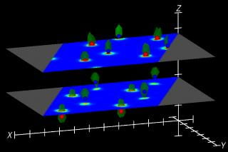
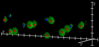
| GaAs(111) (2×2) | O/Cu(104) |
 |
 |
| Sb/Ge(113) Work in progress | O/Cu(104) Work on Exp. Data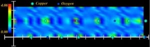 |
Experimentally measurable are the moduli |F(h,k,l)| of the structure factors of the 2D unit cells, with discrete Miller indices h and k specifying the reciprocal-space rods, and reflecting the periodicity parallel to the surface. The index l is continuous along each rod, a consequence of the lack of periodicity perpendicular to the surface.
Determination of the structure of the surface requires a knowledge of the phases of these structure factors, which are not directly avaliable from the experiment. Information about these phases is available in coded form in the diffraction data, but coaxing it out into a usable form is a problem that has defied solution up to now.
In this page and the ones clickable from here, we show the results of an algorithm that combines information about the known part of the structure (the bulk) with the measured difraction data to recover the (unknown) electron density distribution of a surface unit cell. In concept this is somewhat similar to holography, where the scattered wave from the known part of the structure plays the role of a reference wave.
Click on the titles of the panels above for more details about the application of the method to the model systems indicated.
THEORY
The algorithm we have developed is called the Phase and Amplitude Recovery And Diffraction Image Generation Method (PARADIGM) [1]. It combines a knowledge of the bulk structure to obtain initial phases of the CTRs and alternate satisfaction of constraints in real and reciprocal space [2] as in recently developed methods of diffractive imaging.
The method has been extended to deal also with cases where the measured diffraction data may be from mixed surface domains [3].
APPLICATIONS TO EXPERIMENTAL DATA
The method has been successfully applied to experimental data in the recovery of the known surface structure of Au(110)-(2×1) including not only the gross feature of the missing row, but even exquisite details of the buckling and relaxations of deeper atomic layers [4]. It has also now been used to determine the previously unknown structures of (2×2)Sb/Au(110) [5] and (rt3xrt3)R30Sb/Au(110) [1].
REFERENCES
- R. Fung, V. L. Shneerson, P. F. Lyman, S. S. Parihar, H. T. Johnson-Steigelman, and D. K. Saldin, Acta Cryst. A63, 239-250 (2007).
- D. K. Saldin, R. J. Harder, V. L. Shneerson, and W. Moritz, J. Phys.: Condens. Matter 13, 10689-10707 (2001).
- D. K. Saldin, R. J. Harder, V. L. Shneerson, amd W. Moritz, J. Phys.: Condens. Matter 14, 4087-4100 (2002).
- P. F. Lyman, V. L. Shneerson, R. Fung, R. J. Harder, E. D. Lu, S. S. Parihar, and D. K. Saldin, Phys. Rev. B 71, art. no. 081402(R) (4 pages) (2005).
- P. F. Lyman, V. L. Shneerson, R. Fung, S. S. Parihar, H. T. Johnson-Steigelman, and D. K. Saldin, Surf. Sci. 600, 424-435 (2006).