[1] S. Lewis, T. Takimato, S. Mehrvar, H. Higuchi, A. Doebley, G. Stokes, N. Sheibani, S. IKeda, M. Ranji, and A. Ideka, “The effect of Tmem135 overexpression on the mouse heart,” PloS one, July 2018.
[2] P. Kadamati, J.J. Sugar, B.J. Quirk, S. Mehrvar, G.G. Chelimsky, H.T. Whelan, T.C. Chelimsky, M. Ranji, “Near-infrared spectroscopy muscle oximetry of patients with postural orthostatic tachycardia syndrome,” Journal of Innovative Optical Health Sciences (JIOHS), July 2018.
[3] Z. Ghanian, S. Mehrvar, N. Jamali, C.M. Sorenson, N.Sheibani, M. Ranji, “Time-lapse microscopy of oxidative stress demonstrates metabolic sensitivity of retinal pericytes under high glucose condition,” Journal of Biophotonics, doi: 10.1002/jbio.201700289, March 2018.
[4] Mette Funding la Cour, S. Mehrvar, J. Heisner, M. Masoudi Motlagh, M. Medhora, M. Ranji, A. Camara, “Optical imaging of whole thorax irradiated rat hearts exposed to ischemia-reperfusion injury,” JBO, Vol. 23, No 1, DOI: 10.1117/1.JBO.23.1.016011, Jan 2018.
[5] S. Mehrvar, M.F. la Cour, M. Medhora, A.K.S. Camara, and M. Ranji, “Time-lapse microscopy of lung endothelial cells under hypoxia,” SPIE BIOS, San Francisco, Feb. 2018.
[6] M. Funding la Cour, S. Mehrvar, J. Kim, M. Zimmerman, J. Hong, M. Ranji, “Optical imaging for the assessment of hepatocyte metabolic state in ischemia and reperfusion injuries,” Journal of Biomedical Optics Express, Vol. 8, No. 10, September 2017.
[7] Z. Ghanian, G.G. Konduri, S.H. Audi, Amadou K.S. Camara and M. Ranji, “Quantitative optical measurement of mitochondrial superoxide dynamics in pulmonary artery endothelial cells,” Journal of Innovative Optical Health Sciences (JIOHS), Vol. 11, No. 1, June 2017.
[8] E. Aboualizadeh, M. Ranji, C.M. Sorenson, R. Sepehr, N. Sheibani, C. Hirschmugl, “Retinal oxidative stress at the onset of diabetes determined by synchrotron FTIR widefield imaging: Towards diabetes Pathogenesis,” Analyst, Feb. 2017.
[9] S. Mehrvar, Z. Ghanian, G. Kondouri, A.K.S. Camara, and M. Ranji, “Time-lapse microscopy of lung endothelial cells under hypoxia,” SPIE BIOS, San Francisco, Feb. 2017.
[10] M. Ranji, M. MasoudiMotlagh, F. Salehpour, R. Sepehr, J.S. Heisner, R.K. Dash, A.K.S. Camara, “Optical Cryoimaging Reveals a Heterogeneous Distribution of Mitochondrial Redox State in ex vivo Guinea Pig Hearts and its Alteration during Ischemia and Reperfusion,” IEEE Journal of Translational Engineering in Health and Medicine (JTEHM), Vol. 4, July 2016. (Featured on IEEE EMBS website)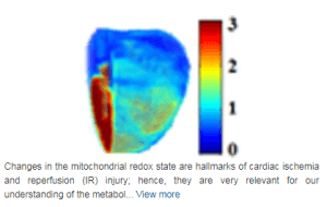
[11] Zahra Ghanian, K. Staniszewski, N. Jamali, R. Sepehr, S. Wang, C.M. Sorenson, N.Sheibani, M. Ranji, “Quantitative Assessment of Retinopathy Using Multi parameter Image Analysis,” Journal of Medical Signals and Sensors, DOI: 10.1109/JTEHM.2016.2570219,Vol. 6, No.2, April 2016.
[12] S.H. Audi, A.V. Clough, S.T. Haworth, M. Medhora, M. Ranji, J.C. Densmore, E.R. Jacobs, “99mTc-Hexamethylpropyleneamine Oxime Imaging for Early Detection of Acute Lung Injury in Rats Exposed to Hyperoxia or Lipopolysaccharide Treatment,” Shock, DOI: 10.1097/SHK.0000000000000605, March 2016.
[13]A.W. Cowley, Jr., C. Yang, N. Zheleznova, A. Staruschenko, T. Kurth, K. Sadovnikov, A. Dayton, M. Hoffman, R. Ryan,M. Skelton, F. Salehpour, M. Ranji, and A. Geurts, “Evidence of the Importance of NOX4 in the Production of Hypertension in Dahl Salt-Sensitive Rats,” Hypertension, DOI:10.1161/HYPERTENSIONAHA.115.06.280, Dec. 2015.
[14] M. MasoudiMotlagh, M. Azimipour, J. Sugar, W.W Linz, G. Michalak, N. Seo, and M. Ranji, “Monitoring hemodynamic changes in stroke-affected muscles using near-infrared spectroscopy,” Journal of Rehabilitation and Assistive Technologies Engineering, Vol. 1, DOI: 10.1177/2055668315614195, Dec. 2015.
[15] F. Salehpour, Z. Ghanian, C. Yang, N.N. Zheleznova, T. Kurth, R. Dash, A.W. Jr Cowley, M. Ranji, “Effects of p67phox on the mitochondrial oxidative state in the kidney of Dahl salt-sensitive rats: Optical fluorescence 3D cryoimaging,” American Journal of Physiology Renal (AJP Rental), Vol. 309, No. 4, F377-F382, DOI: 10.1152/ajprenal.00098.2015, Aug. 2015.
[16] M. MasoudiMotlagh, R. Sepehr, N. Sheibani, C.M. Sorenson, and M. Ranji, “Optical cryoimaging of mitochondrial redox state in Bronchopulmonary-dysplasia injury models in mice lungs,” Quantitative Imaging in Medicine and Surgery, Vol. 5, No.1, Feb. 2015.
[17] Z. Ghanian, A.G. Eis. G.K. Konduri, and M. Ranji, “Optical studies of oxidative stress in pulmonary artery endothelial cells,” SPIE BIOS, San Francisco, Feb. 2015.
[18] F. Salehpour, C. Yang, T. Kurth, A. W. Cowley Jr, and M. Ranji, “Optical cryoimaging of rat kidney and the protective effect of p67 in salt-induced hypertension,” SPIE BIOS, San Francisco, Feb. 2015.
[19] A. W. Cowley Jr., F. Salehpour, C. Yang, T. Kurth, M. Ranji, “Cryofluorescence 3D imaging shows mutation p67phox improves metabolic function and reduces oxidative stress in the renal medulla of Dahl salt-sensitive rats,” American Heart Association Conference, High Blood Pressure Research, San Francisco, CA, Sept. 2014.
[20] T. R. Schaid A. H. Abdelhafeez, M. Ranji, R. Love, S. Audi, S. Kaul, F. Bashiri, E. MasoudiMotlagh, F. Salehpoor, E. Jacobs, J. C. Densmore, “Surface Fluorescence Studies Of Tissue Mitochondrial Redox State In Ex-vivo Lung Perfusion,”Shock 37th annual Conference, June 2014.
[21] Z. Ghanian, K. Staniszewski, R. Sepehr, C.M. Sorenson, N. Sheibani, and M. Ranji, “Cytometric classification of retinopathic injury,” SPIE Proceeding of SPIE BIOS, San Francisco, Feb. 2014.
[22] W.W. Linz, G. Michalak, M.M. Motlagh, N. Seo, M. Ranji, “Blood volume and oxygenation changes in the skeletal muscles of stroke patients measured using non-invasive surface near-infrared spectroscopy,” SPIE Proceeding of SPIE BIOS, San Francisco, Feb. 2014.
[23] Z. Ghanian, S. Maleki, F. Assadi-Porter and M. Ranji, “Optical imaging of mitochondrial redox state in rodent models with 3-iodothyronamine,” Journal of Experimental Medicine and Biology, Vol. 239, pp. 151- 158, DOI: 10.1002/jbio.201300033, Dec. 2013.
[24] Z. Ghanian, S. Maleki, F. Assadi-Porter and M. Ranji, ” Optical imaging of mitochondrial redox state in rodent models with 3-iodothyronamine,” Journal of Experimental Medicine and Biology, Vol. 239, pp. 151- 158, DOI 10.1002/jbio.201300033, Dec. 2013.
[25] R. Sepehr, S. Audi, K. Staniszewski, E. R. Jacobs, and M. Ranji, “Novel Flurometric Tool to Assess Mitochondrial Redox State of Isolated Perfused Rat Lungs after Exposure to Hyperoxia,” IEEE Journal of Translational Engineering in Health and Medicine, Vol. 1, No. 1, 1500210-1_1500210-10, Nov. 2013.
[26] Z. Ghanian, M. MasoudiMotlagh, S. Maleki, Z. Bolandnazar, F. Assadi-Porter, M. Ranji, ” Optical Redox Imaging of Metabolic Dysfunction in Polycystic Ovary Syndrome,” BMES Annual conference, Seattle, Sept. 2013.
[27] Z. Ghanian, S. Maleki, C.M. Sorenson, SunYoung Park, N. Sheibani and M. Ranji, “Organ specific optical imaging of mitochondrial redox state in a rodent model of hereditary hemorrhagic telangiectasia-1,” Journal of Biophotonics, Vol. 6, No. 6, June 2013.
[28] R. Sepehr, S. Audi, S. Maleki, A.L. Eis, G.G. Konduri, M. Ranji, “Optical Imaging of Lipopolysaccharide-induced Oxidative Stress in Acute Lung Injury from Hyperoxia and Sepsis,” Journal of Innovative Optical Health Sciences (JIOHS), Vol. 6, No.3 June 2013.
[29] W. Linz, G. Michalak, M. MasoudiMotlagh, N. Seo, M. Ranji, “Hand function assessment of stroke patients using non-invasive surface near-infrared spectroscopy,” Great Lake Biomedical Conference, GE Healthcare, Milwaukee, April 2013 (awarded third place).
[30] M. MasoudiMotlagh, S. Maleki, F. Assadi-Porter, M. Ranji, “Optical Redox Imaging to Monitor Metabolic Dysfunction in Polycystic Ovary Syndrome,” 2013 Great Lake Biomedical Conference, GE healthcare, Milwaukee, April 2013.
[31] Z. Ghanian, S. Maleki, C.M. Sorenson, N. Sheibani, M. Ranji, “Optical Imaging of Cellular Redox State Related to Retinopathy Dysfunction in Endoglin Heterozygous (Eng+/-) Mice,” UWM CEAS Research Day, April 2013.
[32] R. Sepehr, K. Staniszewski, E. R. Jacobs, S. Audi, and M. Ranji, “Fluorometry of ischemia reperfused rat lungs in vivo.”Proceeding of SPIE BIOS, San Francisco, Jan. 2013.
[33] Z. Ghanian, S. Maleki, S. Gopalakrishnan, R. Sepehr, J. T. Eells and M. Ranji, “Optical imaging of oxidative stress in rodent model of retinitis pigmentosa.” Proceeding of SPIE BIOS, San Francisco, Jan. 2013.
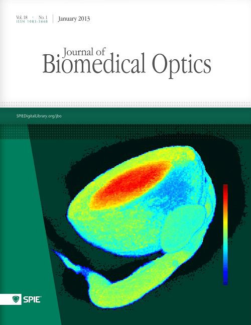
[34] Sepideh Maleki, Sandeep Gopalakrishnan, Reyhaneh Sepehr, Zahra Ghanian, Heather Schmitt, Janis Eells, Mahsa Ranji, “Optical imaging of tissue mitochondrial redox state in rodent model of retinitis pigmentosa.” Journal of Biomedical Optics, Vol. 18, No. 1, Jan 2013.(Selected as cover page).
[35] Kevin Staniszewski, Said H. Audi, Reyhaneh Sepehr, Elizabeth R. Jacobs, and Mahsa Ranji, “Surface Fluorescence Studies of Tissue Mitochondrial Redox State in Isolated Perfused Rat Lungs.” Annals of Biomedical Engineering, Vol. 41, Jan. 2013.
[36] Reyhaneh Sepehr, Kevin Staniszewski, Sepideh Maleki, Elizabeth R. Jacobs, Said Audi, Mahsa Ranji, “Optical imaging of tissue mitochondrial redox state in intact rat lungs in two models of pulmonary oxidative stress.” Journal of Biomedical Optics, Vol. 17, No. 4, April 2012.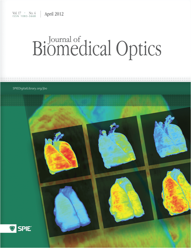
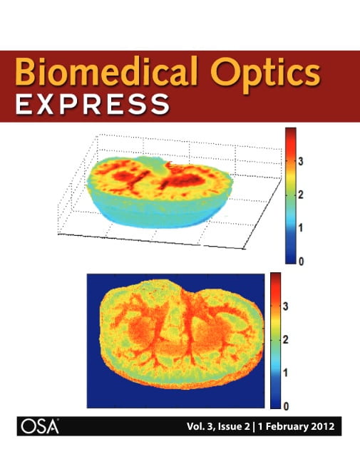
[37] Sepideh Maleki, Reyhaneh Sepehr, Kevin Staniszewski, Nader Sheibani, Christine M. Sorenson, and Mahsa Ranji, “Mitochondrial redox studies of oxidative stress in kidneys from diabetic mice.” Journal of Biomedical Optics Express, Biomedical Optics Express, Vol. 3, No. 2, pp. 273-281, Feb. 2012 (Selected as cover page).
[38] Reyhaneh Sepehr, Sepideh Maleki, Kevin Staniszewski, Said Audi, Mahsa Ranji, “Optical Studies of Mitochondrial Redox State in Normoxic and Hyperoxic Rat Lung Tissue.” SPIE Photonics West proceeding, San Francisco 2012.
[39] S Maleki, R. Sepehr, K. Staniszewski, N Sheibani, C.M. Sorenson, M. Ranji, “Optical Cryoimaging of Kidney Mitochondrial Redox State during Diabetes.” SPIE Photonics West proceeding, San Francisco 2012.
[40] Kevin Staniszewski, Reyhaneh Sepehr, Christine M. Sorenson, Nader Sheibani, Mahsa Ranji, “Quantification and Classification of Retinopathic Injury Using Image Cytometry and Vasculature Feature Extraction.” SPIE Photonics West proceeding, San Francisco 2012.
[41] Reyhaneh Sepehr, Kevin Staniszewski, Sepideh Maleki, Nader Sheibani, Christine Sorenson, Mahsa Ranji, “Optical Cryoimaging of Kidney Mitochnodrial Redox States and the Effect of BCL-2 Family Expression.” Proc. BMES, Hartford, 2011.
[42] R. Sepehr, S. Audi, K. Staniszewski, S Maleki, M. Ranji, “Fluorescence Spectroscopy and Cryoimaging of Rat Lung Tissue Mitochondrial Redox State.” Proc. SPIE 8087, 80870A, Munich, 2011.
[43] Muneaki Matsubara, Mahsa Ranji, Bradley G. Leshnower, Mio Noma, Sarah J Ratcliffe, and Britton Chance, Robert C. Gorman, Joseph H. Gorman III, “The Influence of Cyclosporine on Mitochondrial Function during Myocardial Ischemia and Reperfusion,” Annals of Thoracic Surgery, vol. 89, pp. 1532-7, May 2010.
[44] Mahsa Ranji, Shoko Nioka, Lin Z. Li, He N. Xu, Dwight L. Jaggard and Britton Chance, “Fluorescent Images of Mitochondrial Redox States of in Situ Mouse Hypoxic Ischemic Intestines,” Journal of Innovative Optical Health Sciences (JIOHS), Vol. 2, No. 4, pp. 365–374, Dec. 2009.
[45] Lin Z. Li, He N. Xu, Mahsa Ranji, Shoko Nioka, and Britton Chance, “Mitochondrial Redox Imaging for Cancer Diagnosis and Therapy,” Journal of Innovative Optical Health Sciences (JIOHS), Vol. 2, No. 4, pp. 325-341, Dec. 2009.
[46] Mahsa Ranji, Diego Calzolari, Ramses Agustin, Jeffrey H. Price, “Quantitative analysis of 3D Fluorescence Images,” Asilomar Workshop on Cytometry, Oct. 2009.
[47] Mahsa Ranji, Muneaki Matsubara, Bradley G. Leshnower, Robin Hinmon, Dwight L. Jaggard, and Britton Chance, Robert C. Gorman, Joseph H. Gorman III, “Quantifying Acute Myocardial Injury Using Ratiometric Fluorometry,” IEEE Transaction on Biomedical Engineering (TBME), Vol. 56, No.6, pp. 1556-1563, May 2009.
[48] Mahsa Ranji, Diego Calzolari, Natalie Prigozhina, Karen A. Wei, Mark Mercola, Jeffrey H. Price, “Automated Tracking of Migration and Diffrentation of Fluorescently Labeled Human Embryonic Stem Cells,” Proc. SPIE Photonics west, Jan 2009.
[49] M. Ranji, D. Calzolari, J.H. Price, “Quantitative analysis of deconvolution methods for fluorescence microscopy images,” Invited, Asilomare Image Cytometry Workshop, Oct. 2008.
[50] M. Ranji, M. Matsubara, B.G. Leshnower, R.Hinmon, D.L. Jaggard and B.Chance, R.C. Gorman and J. H. Gorman III, “Optical Biopsy of Apoptosis in Ischemic Myocardium with Fluorometry,” Proc. of AHA (American Heart Association), Nov. 2007.
[51] M. Ranji, M. Matsubara, M. Grosso, D.L. Jaggard and B.Chance, R.C. Gorman and J. H. Gorman III, “Fluorescence spectroscopy to assess apoptosis in myocardium,” Proc. of SPIE, Vol. 6438, pp. 64380J1- 64380J4, Feb. 2007.
[52] Mahsa Ranji, Shinya Kanemoto, Muneaki Matsubara, Michael A. Grosso, Joseph H. Gorman III, Robert C. Gorman, Dwight L. Jaggad and Britton Chance, “Fluorescence spectroscopy and imaging of myocardial apoptosis,” Journal of Biomedical Optics, Vol. 11, No. 6, Dec. 2006.
[53] Mahsa Ranji, Dwight L. Jaggard, Sofia V. Apreleva, Sergei Vinogradov, and Britton Chance, “Simultaneous fluorometery and phosphorometry of Langendorff perfused rat heart: ex vivo animal studies,” Optics Letters, Vol. 31, Issue 20, pp. 2995-2997, Oct. 2006.
[54] M. Ranji, D.L. Jaggard and B.Chance, “Observation of mitochondrial morphology and biochemistry changes undergoing apoptosis by angularly resolved light scattering and cryoimaging,” Proceedings of SPIE, Vol. 6087, pp. 60870K1 – 60870K9, Jan. 2006.