Thermoacoustic computerized tomography (TCT) is a hybrid imaging technique. Ideally, electromagnetic (EM) energy is deposited impulsively in time and uniformly throughout the imaging object, causing thermal expansion. Rapid thermal expansion generates thermoacoustic signals which are detected by ultrasound receivers placed outside the field of view. Different types of tissue absorb different amounts of EM energy, which leads to variable heating and thermal expansion throughout the object. By “listening” for the emitted thermoacoustic pulses from different locations around the object, we collect a TCT dataset (sinogram) similar to those collected by x-ray CT scanners. Dr. Patch has developed an inversion formula for idealized TCT data and now works to account for physical and experimental effects upon TCT data. Her long-term goal is to quantify the robustness of TCT across different sizes, depths, and types of cancer.
The Patch Lab utilizes very high frequency (VHF) EM energy, to enable whole organ imaging. Choice of EM frequency impacts the contrast mechanism and imaging depth. For instance, optical energy is preferentially absorbed by hemoglobin in blood and has a penetration depth of less than 2 cm in soft tissue. Microwaves heat pure (deionized) water and can develop standing waves in tissue creating “hotspots” and also “coldspots” from which no thermoacoustic information is available. (Think of uneven heating in microwave ovens without rotary tables.) Very high frequency energy is absorbed by ions which are present in electrically conductive blood and other fluids. VHF is used to excite MRI signals and can easily penetrate the abdomen of a large adult, enabling whole organ imaging.
Prostate cancer is notoriously difficult to image so Dr. Patch’s lab has imaged fresh human prostates immediately after surgery using VHF-induced TCT. Prostatic fluids produced by healthy prostates have high concentrations of citrate and other ions, such as zinc, so electrical conductivity is greater than that of blood and human plasma. However, ion concentrations vary by zonal area of the prostate. In the normal peripheral zone, which is the most commons site of prostate cancers, citrate levels are high, but in the presence of prostate cancer, these levels decrease which in turn decreases electrical conductivity. Conversely, it is well established that zinc and citrate levels are lower in normal central zone tissue, but elevated in BPH, which typically occurs in the central zone. In a pilot study (PatchHullThomasGriepJacobsohnSeePMB15), preliminary thermoacoustic images appear to corroborate these findings. Reconstructions frequently visualize the verumontanum, which is used as an anatomical landmark to compare thermoacoustic images to histology slides. In the figure below, the posterior region is subdivided into regions of interest corresponding to tissue type as annotated on histology slides. Mean image values in cancer-free regions are four times greater than in regions with extensive disease.
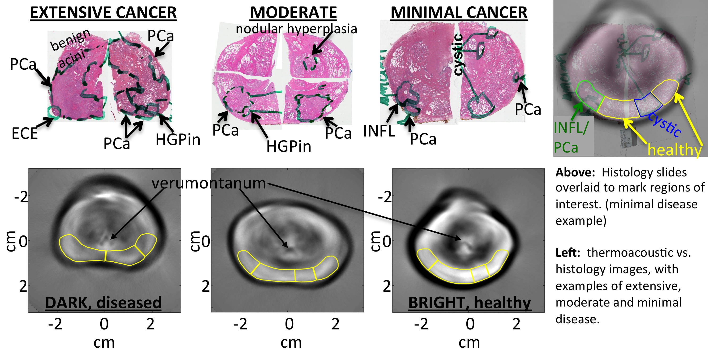 Acronyms: PCa~prostate cancer, INFL~inflammation, HGPin~high-grade prostatic intraepithelial neoplasia, ECE~extracapsular extension of cancer outside the prostate
If results of a larger study also support the hypothesis that VHF-induced thermoacoustic imaging is sensitive to prostate cancer then development of a clinical prototype for in vivo imaging will be warranted.