(Research supported by the Natural Environmental Research Council of the UK, and by joint British Council/Research Council of Norway Funds)
1) Do species other than D. tertiolecta show this cell death response? Do other factors trigger it?
(work assisted by Honours student Pauline McIntyre)
We have surveyed 16 species and we are currently working with 8 more.
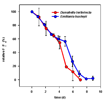
Judged by variable fluorescence emissions (an index of photosynthetic ability), we have found that generalisations do not apply across similar taxa. There are some species that show a similar responses to D. tertiolecta:

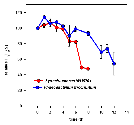
Some species show a more gradual decline and eventually come to a stable level without substantial cell losses:

Some species just don’t seem to care about being in the dark:

So far, other stresses applied (e.g. deprivation of nitrogen and phosphorus, heat shock) have not resulted in cell death in any species. However, the process does appear to occur faster at elevated temperature (6 days at 17 degrees versus 2-3 days at 25 degrees).
2) Do any cells survive the decline? What happens when the cells are put back in the light?
(work assisted by Honours student Michael Scott)
Using Evans Blue (which stains only dead cells), we have followed the kinetics of the cell decline in darkness, and recovery in light.

Some cells do survive, but there is a substantial delay before they can recover when put back into the light. As well, we have not merely selected out certain suceptible cells…the culture decline can be reproduced in darkness again.
3) Could viruses be involved in this process?
Marine viruses that attack and lyse phytoplankton are currently an active topic of research. We have argued previously that this phenomenon has differences from typical viral attack, notably an apparent independence on cell or innoculum density.
Nonetheless, we are pursuing several lines of work.
(collaborations with Drs. G. Bratbak and C. Brussaard, Dept. of Microbiology, University of Bergen, Noway)
a) Are there viruses in the Dunaliella tertiolecta cultures?
b) Is there evidence of protease expression during viral cell lysis of other species?
(collaboration with Dr. W. Wilson, Marine Biological Association of the UK, Plymouth)
c) Could there be a temperate virus (i.e one hidden in Dunaliella tertiolecta’s DNA and activated by specific triggers)?
4) Is this really a case of apoptosis?
(collaborations with Dr. A. Wallace, School of Biology and Biochemistry, Queen’s University; work assisted by Honours student Michael Scott)

The photograph shows an agarose gel of DNA, stained with ethidium bromide. The first lane contains molecular size markers, the next three lanes are samples of Dunaliella tertiolecta DNA taken at various stages of culture decline, and the last lane is DNA from Dunaliella that has been randomly degraded.
Note that in lane 2 there are discrete fragments of DNA i.e. not just one big piece, indicating the whole genome, and not a smear (as in lane 5) indicating random degradation. Such ‘laddering’ of DNA is taken as evidence of apoptosis.
We are currently trying to support this by: